How it Works
SonoSim, The Easiest Way to Learn Ultrasonography®, uses a proven structure and method for learning ultrasound that combines:
- A Didactic, Cloud-Based, Multimedia Course
- Real-Time Knowledge Checks & a Mastery Test, &
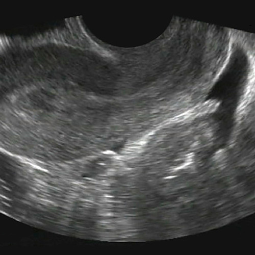
- A Series of Hands-On, Real Patient Scanning Cases Accessed with the SonoSimulator® Ultrasound Probe
CME*, a Certificate of Completion, an ultrasound image portfolio come with your module(s).
The SonoSimulator Ultrasound Probe is a one-time purchase and is your access key to the SonoSim modules you license annually. License to each module is purchased in the first year, and maintained with an annual membership fee. Keep scanning and maintain all your member benefits, including annual access to all your modules, with your low annual membership fee of $295 (complimentary in your first year).


