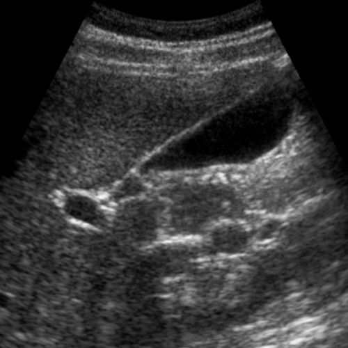
Liver Anatomy & Physiology Ultrasound Training

Learn how to use ultrasound in the evaluation of the liver, including anatomy and physiology, and sonographic anatomy of the liver. Optimal transducer selection, patient positioning, imaging approaches, and techniques for sonographically evaluating the liver are discussed.
CME Credits:
2.75
Cases:
3
Price US$399.00
Adding to cart… The item has been added

How it Works

Each SonoSim module contains:

  • A Didactic, Cloud-Based, Multimedia Course
  • Real-Time Knowledge Checks & a Mastery Test
  • A Series of Hands-On, Real Patient Scanning Cases Accessed with the SonoSimulator® Ultrasound Probe

The SonoSimulator Ultrasound Probe is a one-time purchase and is your access key to the SonoSim modules you license annually. License to each module is purchased in the first year, and maintained with an annual membership fee. Keep scanning and maintain all your member benefits, including annual access to all your modules, with your low annual membership fee of $295 (complimentary in your first year).

Course Highlights

SonoSim cloud-based, multimedia, didactic courses are created by leading ultrasound experts, and are internet accessible immediately after purchase, on any device. There are knowledge checks with real-time feedback throughout each course, as well as a Mastery Test that is automatically graded.

The course in this module covers topics including:

  • Liver Anatomy including Portal Triad & Porta Hepatis
  • Liver Physiology
  • Sonographic Anatomy of the Liver, including the Various Lobes and Planes
  • Sonographic Techniques, including the Use of Doppler Mode
  • Pearls & Pitfalls when Evaluating the Liver

SonoSimulator Scanning Cases

The SonoSimulator® helps develop and maintain the critical visuomotor and visuospatial skills that are central to image acquisition and interpretation with real patient imagery, expert tutorials on-demand, and real-time feedback on success.

The 3 real patient scanning cases in this module include and cover:

  • Patient Types: Male & Female, 2 Adult & 1 Pediatric
  • Relevant Findings/Pathology: Normal Examples of Liver Anatomy
  • Transducer Types: Curvilinear
  • Special Case Features: Additional Abdominal CT Imaging, Anatomic Layer Removal

Continued Medical Education

The utility of ultrasound for evaluating the liver continues to evolve in concert with improved image resolution provided by modern-day ultrasound machines, advances in Doppler-mode imaging, and scanning protocols that integrate ultrasound contrast. Liver ultrasound can be used to evaluate a multitude of conditions, such as liver cirrhosis, liver failure, portal hypertension, tumors, abscesses, and traumatic injury. The Liver: Anatomy & Physiology Module begins with an overview of the role of ultrasound in the evaluation of the liver. A brief review of liver anatomy and physiology is provided. The sonographic anatomy of the liver and classification systems of the liver are described. Optimal transducer selection, patient positioning, imaging approaches, and techniques for sonographically evaluating the liver are discussed. The course concludes with imaging tips and pitfalls and a summary of salient teaching points.

This activity has been planned and implemented in accordance with the accreditation requirements and policies of the Accreditation Council for Continuing Medical Education (ACCME) through the joint providership of the American College of Emergency Physicians, the Illinois College of Emergency Physicians, and SonoSim, Inc. The American College of Emergency Physicians is accredited by the ACCME to provide continuing medical education for physicians.

The American College of Emergency Physicians designates this enduring material for a maximum of 2.75 AMA PRA Category 1 Credits™. Physicians should claim only the credit commensurate with the extent of their participation in the activity.

Approved by the American College of Emergency Physicians for a maximum of 2.75 hours of ACEP Category I credit.